Self Muscle Massage- pt 16 Forearm/Wrist
This is part sixteen in the Self Muscle Massage Series. In the introduction post to this series we introduced and demonstrated the three muscle release techniques that will be used in this post. If you would like to review them, click here. If you would like to see any other parts of the series, click here.
In this installment of the series we’re going to be moving from the elbow down into the forearm, wrist and hand. Typically, as you move further away from the core of the body, the muscles get smaller and become more prone to injury through repetitive overuse situations. It is also why bony injuries such as fractures and joint dislocations become more common where the muscles are unable to counteract the full load of the body in a fall situation onto the arm. In this area, the most common muscular injuries are lateral and medial epicondylitis (also known as tennis and golfers elbow).
Anatomy
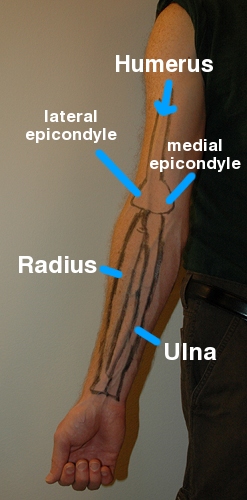
Bony Landmarks
As you may recall from the last post, the elbow moves in four ways- 1) bending (flexion), 2) straightening (extension), 3) pronation (rotating the forearm so that the hand is facing down towards the floor), and 4) supination (rotating the forearm so that the hand is facing up towards the ceiling). With those four motions in mind, an easy way to visualize the elbow joint is to think of it as one bone coming down from the shoulder (this is the humerus). As it’s dangling there, two bones that are already connected to each other (this is the radius and ulna) then literally hook on to that bone. This forms the actual elbow joint and allows you to bend and straighten your arm. Rotation at the elbow actually occurs because of the two lower bones and how they are connected to each other. The rotation of the forearm occurs because the radius is able to rotate over the ulna (it is connected directly to the humerus and provides the bending/straightening).
In addition to the elbow, we now have to add in movement at the wrist itself. It also moves in four ways. With your palm facing the floor, these movements are: 1) extension (bending the wrist up towards the back your hand), 2) flexion (bending the wrist down towards the palm of your hand), 3) ulnar deviation (bending the wrist towards your pinky finger), and 4) radial deviation (bending the wrist towards your thumb).
#1 Humerus. The humerus is the long bone of the arm that moves down from the shoulder to the elbow. As it moves down the arm, the narrow bone becomes wider at the elbow. If you cup your hand under your elbow you will feel the two “knobs” on either side. These are called the epicondyles (the medial is on the inside closest to the body, and the lateral is on the outside away from the body). The epicondyles are important! These are the two main attachment points for the most of the muscles in the forearm. The epicondyle closest to your side is the called the medial epicondyle. This is where the wrist flexors attach (meaning the muscles that bend your wrist up towards your palm). Strains/sprains/tendonitis in this area is commonly called golfers elbow or medial epicondylitis. The “knob” on the outside is your lateral epicondyle. This is where the wrist extensors attach (meaning the muscles that bend your wrist up towards the back of your hand). Sprains/strains/tendonitis in this area is commonly called tennis elbow or lateral epicondylitis.
#2 Forearm bones- Radius and Ulna. These two bones connect in two spots (up near the elbow and then again down at the wrist). The ulna actually hooks onto the humerus at the back of the elbow. This part of the ulna is known as the olecranon. The second forearm bone, the radius, then connects to the ulna. Together, these two bones rotate to produce supination and pronation of the arm. Without these you would be unable to turn keys, door knobs etc. An easy way to differentiate which bone is which is to look at your hand. The bone on the side of your thumb is the radius, while the bone on the side of your pink is the ulna. As you rotate your arm back and forth you can see how the radius rotates over the ulna which is connected to the humerus.
#3 The wrist + hand. Instead of rambling on about all of the tiny bones that makes up the hand and wrist, I’m going to stick with a simple overview. Just below the radius and ulna are the carpal bones. There are eight of them and they are arranged like little rocks between the long bones of the forearm and those of the fingers and thumb. These small bones allow for wrist movement while the long finger bones allow for grip, pinch, etc. The bones of the fingers and thumb are arranged in segments to allow for increased mobility. In order they are- 1) from the wrist to your knuckles are the metacarpals and 2) from your knuckles to your finger tips are the phalanges. As you look at your fingers, there is a little bone on either side of the creases (where the finger bends). Your index finger for example has two creases and therefore three phalanges while your thumb only as one crease and two phalanges.
Muscles
When it comes to the wrist and hand, an easy way to think of the muscles is to think about what fingers of the hand they control (they are traditionally referred to as digits 1-5 with 1 being the thumb and 5 being the pinky or little finger). With this in mind, the muscles will either control the thumb (digit #1), little finger (digit #5), or the three fingers in between (index, middle, ring or digits 2-4).
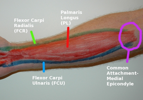
#1 Wrist Flexors Group. There are three separate muscles in the wrist flexor group and they all start on the medial epicondyle of the humerus before moving down the arm to the wrist. As I stated above, the hand and fingers can be broken down into three segments (thumb, middle fingers, little finger). You can use those segments to follow the muscles. The small blue muscle is the flexor carpi ulnaris and it travels from the epicondyle, over the wrist joint to insert on the 5th digit. The middle red muscle is the palmaris longus and it crosses the wrist joint to insert on digits 2-4. The green muscle is the Flexor carpi radialis and it attaches to the base of digits 2+3. Together all three of these muscles are responsible for bending the wrist down towards the palm of your hand. The green and blue muscles are also able to assist with radial and ulnar deviation (bending the wrist towards the thumb and towards the pinky finger respectively). If you bend your wrist up towards your palm, you will see the three tendons pop up at the wrist. Likewise, you place your thumb on the medial epicondyle and bend your wrist back and forth, you will be able to feel the common tendon move.
#2 Wrist Extensors Group. Like the flexor group, the extensor group contains multiple individual muscles that all share a common attachment at the elbow. For this group the muscles all begin on the lateral epicondyle and then move down the back of the forearm to cross the wrist joint before inserting on the carpals and metacarpals. In this group you can again use the three segments of thumb, middle fingers and little finger. This gives us the Extensor carpi ulnaris (to the the pinky finger), the extensor digitorum (to digits 2-5) and extensor carpi radialis (to digits 2+3). Due to their attachments on the end, the ECU is able to bend the wrist towards the pinky and the ECR is able to bend the wrist towards the thumb. If you bend your wrist up towards the back of your hand, you will see the three tendons pop up at the wrist. Likewise, if you place your thumb on the lateral epicondyle and bend your wrist back and forth, you will be able to feel the common tendon move.
Soft Tissue Release
What you’ll need: stick/foam roller and tennis ball
The techniques: click here for an introduction to the techniques and a video demonstration
1) Lengthening/elongation with the foam roller or stick.
2) Cross friction with your hand or tennis ball.
3) Sustained pressure or trigger point release with the tennis ball.
Key Areas to Work On
#1 Foam Roller. When working on the forearm, start by loosening up the larger and more superficial muscles with the foam roller. For this area you’ll want to use a raised surface for a few reasons: 1) the muscles are small and full pressure will likely be uncomfortable due to how superficial the bones are, and 2) this area is super easy to work on- just rotate the palm up or palm down and you’ll quick access to both sides. A bed or table works best (for example- I use my coffee table). Just like the other areas of the body, shoot for 3-5 minutes before moving to the deeper techniques.
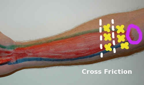
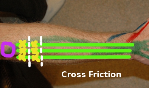
#2 Tennis Ball- Cross Friction. The key with cross friction is to remember that you are working perpendicular to the muscle fibers. This means that you will be working in a side to side (horizontal) direction when working on the forearm arm. The movement itself is very small (maybe 1-2 inches). Sink the tennis ball in deep, relax and then maintain that depth as you work. If you feel like the ball or your fingers are rolling or sliding, you’re moving too much. When working on the forearm, the primary locations for cross friction work will be on the common tendons of the flexor and extensor muscle groups where they insert onto the epicondyles. Sitting with the tennis ball will work best for the flexor group. The extensor group is easiest using your fingers/thumb versus the tennis ball. See the video for further details. If you’re still unsure of the cross friction technique and how to properly do it, click here for a review.
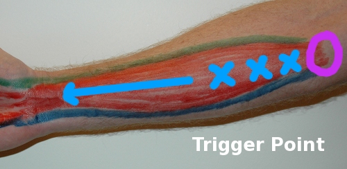
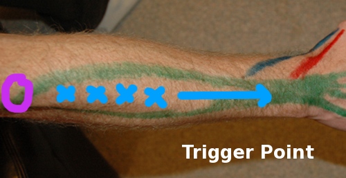
Key areas- (the purple circles represent the epicondyles in the pictures above!)
1) Wrist extensor tendon. Use the palpation guidance above to find the common tendon with your thumb. Then slide your fingers down approx one inch and your index/point finger. From here you can work each of the three bands of the muscle group (ECR, ED, ECU). After you work on all three, then slide down an inch and repeat. This can be done for the entire length of the muscle group. See the video for further details.
2) Wrist flexor tendon. Use the palpation guidance above to find the common tendon and position the tennis ball just below it. From here you can work each of the three bands of the muscle group (FCR, PL, FCU). After you work on all three, then slide down an inch and repeat. This can be done for the entire length of the muscle group. See the video for further details.
#3 Tennis Ball- Trigger Point.
When moving onto trigger point areas, remember, let the tennis ball sink in nice and deep and just sit on it. If after 2-3 minutes it hasn’t released, move onto the next spot!
Key Areas: The purple circles are the epicondyles!
1) Muscle bellies of the wrist flexors and extensors. For the extensors, use your thumb and sink right into the thickest part of the muscle. HOLD it there. This can also be done standing up with the ball on the wall. For the flexors, use the tennis ball while sitting. For trigger point, you’ll want to stay closer to the middle of the group due to the small size of the muscles. Trigger point works best on the thicker parts of the muscles (aka the muscle bellies). As you move closer to the wrist, you’ll be working with a smaller and smaller area. With that in mind, stick to the middle and work your way down! See video for further details.
Video
Here is a video demonstration for self muscle release for the forearm and wrist using a foam roller and tennis ball.
References
1) Hammer, Warren. (2007). Functional Soft-Tissue Examination and Treatment by Manual Methods, 3rd edition. Jones and Bartlett Publishers, Inc, Sudbury, MA.
2) Hyde, Thomas and Gengenbach, Marianne. (2007). Conservative Management of Sports Injuries, 2nd edition. Jones and Bartlett Publishers, Inc, Sudbury, MA.
3) Moore, Keith and Dalley, Arthur. (1999). Clinically Oriented Anatomy, 4th edition. Lippincott Williams and Wilkins, Baltimore, MD.
4) Muscolino, Joseph. (2009). The Muscle and Bone Palpation Manual. Mosby, Inc, St. Louis, MO.

