Self Muscle Massage- pt 5 Anterior Hip
This is the fifth part of the Self Muscle Massage series. In the introduction post to this series, we discussed and demonstrated the three soft tissue release techniques that will be used below. If you missed it or would like to review them, click here. If you would review the other muscle groups covered so far, click here.
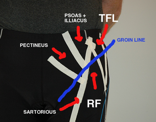
In this installment of the series we’re going to be finishing up with the hip by covering the front or anterior portion. This area includes your hip flexors (psoas and iliacus), sartorious, TFL, rectus femoris (large quad muscle), and pectineus/obturator externus. The front of the hip is important in pulling the leg forward and lifting the femur to allow for clearance of the entire lower extremity (i.e. up stairs, inclines, etc). Like the other sections of the hip, the anterior aspect is also prone to injury. It is a common site of muscle contractures (chronic loss of flexibility) and musculotendinous injuries (i.e. tendonitis and “snapping hip” syndrome).
Potential Causes of Injury
1) During normal ambulation, the hip flexors are active during the swing phase (meaning they pull the leg through to start the stance phase all over). With normal push off at the big toe, knee, and hip, part of this motion is passive (meaning the hip flexor can use that momentum to lesson the load on itself). If the push off is incomplete ( as is the case with chronically tight gastrocs and hamstrings for example), the hip flexors must pick up the slack and actively pull the leg through. This can result in a tendon/muscle overuse injury or an acute injury due to a forceful contraction.
2) Due to their location, the muscles in the front of the hip are prone to contractures due to prolonged sitting. As the muscles adapt to that shortened position, they limit hip extension and subsequently push off. This can result in tendon/muscle injuries.
3) In the presence of an anterior pelvic tilt (when the front of the pelvis tips down towards the ground), the anterior muscles are kept in a shortened position and can lead to contracture over time. The hamstring muscles in the back of the thigh are then weakened by being kept in a lengthened position. As the back of the hip is unable to extend, the hamstrings are also unable to compensate, and the hip flexors are left to do all of the work.
4) Combine any of the above scenarios with a deviation from mid-line (when the the whole leg is pulled in towards mid-line) and it is not uncommon for the tight and overworked anterior muscles to get pulled out of their grooves and begin sliding/snapping over the hip bones. This is commonly referred to as snapping hip syndrome.
Anatomy:
Landmarks
In the front of the hip there are two landmarks used to navigate the muscles. One is bony and the other is soft tissue.
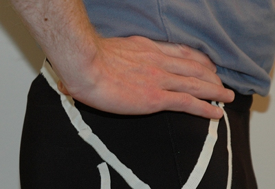
#1 The ASIS (anterior superior iliac spine)- while the PSIS is at one end point of the iliac crest towards the back, the ASIS is the end point towards the front. To find it, start with your hands on your hips and your fingertips pointing towards your stomach. Unlike the other two bony landmarks that are small bumps, the ASIS are larger and easily palpable when you follow the iliac crest forward. Visually, when you lay on your back, they are the two hip bones sticking out towards the ceiling.
#2 The groin line- this is a visible soft tissue landmark. When you flex your hip (lift your knee up towards the ceiling), it is the line/crease between your thigh and pelvis.
Muscles
#1) The TFL- the TFL is a small muscle that originates from the ASIS and inserts into the ITB itself. To find this muscle, lay on your back with your hand on the ASIS. With your knee bent, flex your hip (bring your knee towards your chest like you were sitting in a chair) and rotate your whole leg IN (think opposite motion of crossing your ankle over your knee). As you rotate the leg, the knee will move in slightly and the outside of your ankle will come up. You will feel the TFL move under your hand.
#2) The Sartorious- this muscle is a s small rope like muscle that originates on the ASIS and wraps across the thigh to insert just below the inside of the knee. Like the TFL, to find this muscle, bend your knee and flex your hip (bring the knee up towards the ceiling). Rotate your whole leg OUT (like you’re trying to prop your ankle up on your other knee). You will feel the Sartorious move under your hand.
**Note: the TFL and Sartorious form a “V” at the front of the hip. Start with your hand on the ASIS and move down towards the groin line. As you rotate the leg in and out, you will be able to feel both muscles moving and sink your thumb right in between them into the “V”. The floor of the V is where the next muscle is, your rectus femoris (RF).
#3) Rectus Femoris (RF) - the RF is one of the four quadriceps muscles and is responsible for extending the knee. Because it is the only quad muscle to cross the hip joint, it also aids in hip flexion. As stated above, to find this muscle, locate the V and sink down into that groove between the sartorious and TFL. There you will find the RF as it moves towards its insertion point.
#4) Psoas/Illiacus - The Psoas and Illiacus muscles are the large hip flexor muscles. They insert into the front of the femur and then move up into the abdominal cavity. The Illiacus muscle inserts into the inside of the pelvic bone and the psoas move up to insert into the lumbar spine. Due to the deeper location, working on the upper portions of the muscles is difficult, specific due to the presence of internal organs, and should be left to professionals. There are a few ways you can work on them however. First, you can work on them at their distal insertion onto the femur. To do so you will need to work your way down the groin line.
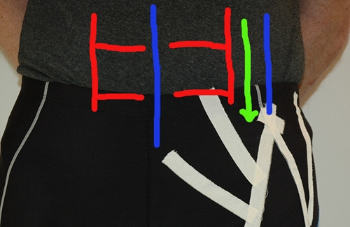
In the picture above, the blue lines represent your two landmarks. The one at midline is your belly button and the other is your ASIS. The red lines represent your abdominal muscles. To find the distal portion of the psoas and illiacus, start on the blue lines and work your fingers in until you find the outer edge of your abs. Move just outside of them (towards the hip) and follow that down to the groin line (this is the green line in the picture).
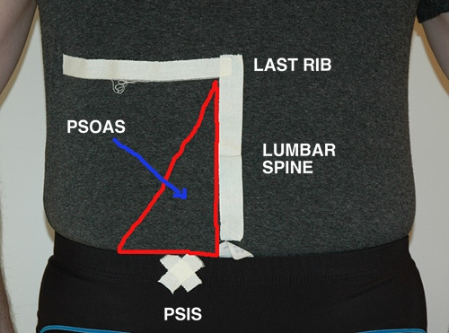
The other area that you can access the psoas is to work on it’s upper insertion into the lumbar spine. To locate the lumbar spine, palpate your last rib and trace it around to your back. This is the level of the last thoracic (midspine) level (T12). Each subsequent bump is the lumbar spine. In the case of the psoas muscle it inserts into levels two through four.
To work on the upper levels, you then want to target just to the side of the lumbar spine. The further away from the spine you move, the less likely your are to be on the right spot.
#5 Pectineus and Obterator Externus- these muscles are furthest down the groin line towards mid-line of the body. Due to their location they work with the adductors to move the leg in to the body. As you find the psoas and iliacus muscles, move medial (towards the mid-line) and feel for a pulse. The femoral artery moves through this area. The muscles are medial (towards mid-line) to the the artery and insert right into the pubic bone. The pectineus is the more superficial and lays directly over the obterator. Note: if you ever feel any numbness/tingling while trying to locate these muscles, move closer to the pubic bone. You’re hitting the femoral nerve.
Soft Tissue Techniques
What you’ll need: foam roller and tennis/trigger point ball.
Techniques:
1) Elongation/lengthening with foam roller.
2) Cross friction with the tennis ball.
3) Sustained pressure/trigger point release with the tennis ball.
Key Area’s to Work On:
1) The trick with the front of the hip is to work the entire groin line from the ASIS to the pubic bone.
2) Due to the deeper nature of the muscles, cross friction and trigger point works best on this area. However, the foam roller can still be used to loosen things up and desensitize the area prior to using the tennis ball.
Video
Here’s a video demonstrating the different techniques for the anterior hip:
References:
1) Moore, Keith and Dalley, Arthur. (1999). Clinically Oriented Anatomy, 4th edition. Lippincott Williams and Wilkins, Baltimore, MD.
2) Hammer, Warren. (2007). Functional Soft-Tissue Examination and Treatment by Manual Methods, 3rd edition. Jones and Bartlett Publishers, Inc, Sudbury, MA.


One Response to “Self Muscle Massage- pt 5 Anterior Hip”