Self Muscle Massage- pt 4 Lateral Hip
This is part four in the self muscle massage series. In the introduction post, we discussed and demonstrated the three soft tissue release techniques that will be used below. If you missed it or would like to review, click here. If you would like to catch up on the other muscle groups we've covered so far, you can do so here.
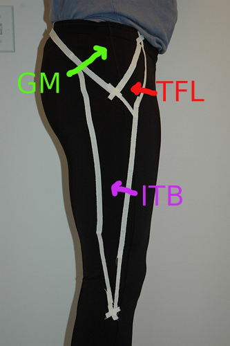
In the next part of our series, we’re going to be talking about the outside or lateral aspect of the hip. This area includes your ITB or Iliotibial Band, your Gluteus Medius/Minimus muscles and your TFL (tensor fascia latae) muscle. The outside of the hip is unique in that it provides stability when standing on one leg. For example, when you step onto your left foot, these structures pull down on your left hip to keep both hips level with each other. If there is injury or weakness, the right side will drop and gait will be compromised as the body then needs to compensate for leg clearance and propulsion. Due to this role in normal gait and trunk stability, the outside of the hip is prone to overuse injuries. In particular, this area is is affected by any alignment abnormalities that move the leg in or out from the vertical mid-line of the body.
Potential causes of injury:
1) On a muscular level, tension is increased on the outside of the hip any time that the knee is pulled in towards the mid-line of the body (sometimes called knock knee or genu valgum). This can happen with chronic tightness (or contracture) of the adductors and internal rotators. When this occurs, the gluteus medius and minimus muscles are held in a lengthened position and subsequently weakened.
2) On a structural level (meaning bones and joints), the knee joint can be pulled in towards the mid-line when there is over-pronation at the foot and ankle. Indirectly, the outside of the hip can also be affected by the presence of an anterior pelvic tilt (when the front of the pelvis rotates down towards the ground). This results in tight hip flexors and weak hamstrings/glutes which alters normal propulsion during gait. The result is commonly that the inner hamstrings and adductors work harder to extend the hip and over time shorten due to the repetitive stress; the knee is pulled in towards the mid-line due to the resulting muscle contracture and the lateral hip muscles overloaded.
Anatomy:
Bony structures
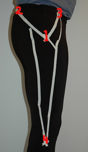
Like the posterior aspect of the hip, working on the outside will require you to know and find a few bony landmarks. There are four: 1) the greater trochanter, 2) the PSIS, 3) the ASIS, and 4) the head of the fibula.
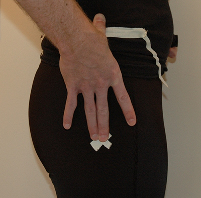
#1 The Greater Trochanter- the GT is a common muscle insertion point on the outside of the hip. To find it, start with your thumb on top of your hip bone at the highest point of the iliac crest. From there, simply lay your hand down over the outside of your hip with your fingers pointed down towards the floor. The GT can be found under or close by where your middle finger is (it will be a small bump).
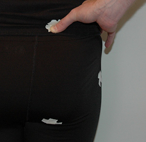
#2 PSIS (posterior superior iliac spine)- To find this one, you’re going to start with your hands on your hip bones (iliac crest) so that your thumb is pointing towards your back and your fingers are pointing forwards towards your stomach. As you reach behind with your thumbs, you’re looking for two small bumps on either side of your spine. Visually, you can see them. They are the two “dimples” at the small of your back.
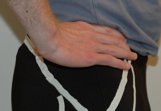
#3 The ASIS (anterior superior iliac spine)- while the PSIS is at one end point of the iliac crest towards the back, the ASIS is the end point towards the front. To find it, start with your hands on your hips and your fingertips pointing towards your stomach. Unlike the other two bony landmarks that are small bumps, the ASIS are larger and easily palpable when you follow the iliac crest forward. Visually, when you lay on your back, they are the two hip bones sticking out towards the ceiling.
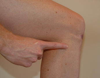
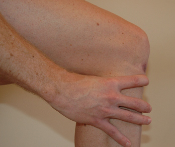
# 4 Gerdy's Tubercle/Fibular Head- I’m including this landmark to point out that your ITB runs all the way down the outside of your leg and inserts BELOW the level of your knee cap. While sitting with your knee bent, wrap your hand around the upper part of your calf so that the space between your thumb and index finger are directly behind the knee and your fingers are wrapped around towards the front of your knee. The fibular head will be the large, bony bump under your index finger. Slide forward just in front of this towards the front of your knee and you will have the insertion point of the ITB, also known as Gerdy's Tubercle. As long as you are working in this area between the knee cap and the fibular head, you're on the right spot.
Soft Tissue Structures
1) GM (gluteus medius + minimus). These two muscles lay one on top of the other and are two fan shaped muscles that originate on the outside of the ilium (hip bone) and insert onto the greater trochanter. The larger medius muscle is the more superficial of the two. Together these muscles abduct the hip (move the leg out to the side and away from vertical midline). They also contribute to rotation of the hip. With the hip in flexion they rotate the knee out (external rotation) and with the hip in extension, they rotate the knee in (internal rotation).
2) TFL (tensor fascia latae). The TFL is a small muscle that originates from the ASIS and inserts into the ITB itself. To find this muscle, lay on your back with your hand on the ASIS. With your knee bent, flex your hip (bring your knee towards your chest like you were sitting in a chair) and rotate your whole leg IN (think opposite motion of crossing your ankle over your knee). As you rotate the leg, the knee will move in slightly and the outside of your ankle will come up. You will feel the TFL move under your hand.
3) ITB (iliotibial band). The ITB starts at the greater trochanter and runs all the way down the outside of the leg to tibia (just in front of the fibular head).
Soft Tissue Techniques
What you’ll need: a foam roller and a tennis/trigger point ball.
Techniques:
1) Elongation/lengthening with the foam roller
2) Cross friction with the tennis ball
3) Sustained pressure/trigger point release with the tennis ball.
Key Areas to Work On:
1) The Gluteus Medius and Minimus take up the whole “fan” on the outside of the hip. Be sure to work the whole fan to get a full release of the muscles. Cross friction and trigger point (sustained pressure) work best on the is area.
2) The TFL is slighly in front of the GM “fan” and below the ASIS. Use the foam roller to release general tension and trigger point any muscle knots or spasms.
3) The ITB is best worked with the foam roller. Use the elongation technique to warm up the area and then cross friction with the roller. Be sure to work from the greater trochanter all the way down below the knee.
Video
Here’s a video to help demonstrate the techniques specific to the lateral hip.
References
1) Moore, Keith and Dalley, Arthur. (1999). Clinically Oriented Anatomy, 4th edition. Lippincott Williams and Wilkins, Baltimore, MD.
2) Hammer, Warren. (2007). Functional Soft-Tissue Examination and Treatment by Manual Methods, 3rd edition. Jones and Bartlett Publishers, Inc, Sudbury, MA.
